
Horses, as flight animals, instinctively remain silent in the face of pain, but a new study, recently published in Equine Veterinary Education, shows that they do in fact have a ‘voice’ if observers are trained to ‘listen’.
The study confirms that if vets are trained to use a Ridden-Horse-Ethogram they are better able to recognise pain-related behaviour in horses, which may reflect lameness or back or sacroiliac pain. This should in turn help them to communicate potential performance problems more effectively with their clients.
Conducted by Dr Sue Dyson, Head of Clinical Orthopaedics at the Centre for Equine Studies at the Animal Health Trust in Newmarket, the study compared the real-time application of the Ridden-Horse-Ethogram with analysis of video recordings of the horses by a trained assessor and determined whether veterinarians, after preliminary training, could apply the ethogram in real time in a consistent way and in agreement with an experienced assessor.
Twenty horse and rider combinations were used for the study. The horses were in regular work and were capable of working ‘on the bit’. They were assessed by a chartered physiotherapist and then a Society of Master Saddlers (SMS) qualified saddle fitter checked the fit, placement, balance and suitability of each horse’s saddle. Eleven horses were found to have ill-fitting saddles and 14 had back muscle tension or pain but these did not influence the behaviour scores.
The horses were also assessed by an independent lameness expert. Sixteen showed low-grade lameness or abnormalities of canter, which were likely to be pain induced but did not prevent the horses from being used for the study.
All 20 horses were given a 15-minute ridden warm-up before executing an 8-minute purpose-designed preliminary level dressage test. During each dressage test a team of 10 equine vets, who were selected from 40 volunteers and given preliminary training, applied the Ridden-Horse-Ethogram. They assessed each horse for the presence of a total of 24 behaviours that occur more commonly in lame horses compared with non-lame horses. It has previously been shown that the presence of ≥8 behavioural markers is likely to indicate the presence of musculoskeletal pain.
All behaviours were scored with a binary method as present or absent. The ethogram was also applied to each horse by an experienced trained assessor (Dr Dyson) and the tests were filmed so that the experienced assessor could make a comparison between her real-time behaviour assessments and video analysis.
There was good agreement between the expert’s scores and the volunteer vets’ scores and excellent consistency in overall agreement among the volunteers. The scores also reflected the volunteers’ capacity to use the ethogram to identify lameness status, with higher scores awarded to the lame horses compared with the non-lame horses.
There was no significant difference in real-time scores and video-based scores for the experienced assessor, verifying the reliability of the system.
“The study confirms that with basic training veterinary observers can use the ridden horse ethogram with consistency as an effective tool to help identify musculoskeletal pain which could reflect lameness or back or sacroiliac pain,” said Dr Dyson. “The volunteers were unanimously positive about the potential value of the ethogram in helping them to determine the presence of musculoskeletal pain in horses performing poorly or at pre-purchase examinations.”
Dr Dyson and her team are now working with US’s popular evidence-based online educational resource Equitopia, in conjunction with Padma Videos, to produce a training video to enable vets, owners, riders, trainers and paraprofessionals to learn how to apply the Ridden-Horse-Ethogram. To find out more email info@equitopiacenter.com
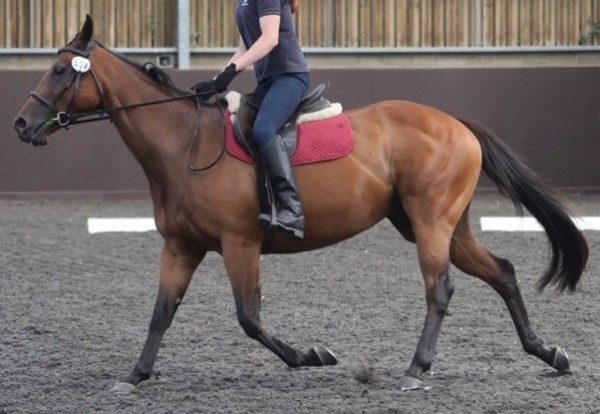
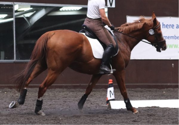
Front of head > 30⁰ in front of the vertical; ears behind vertical for 5 seconds; intense stare;bit pulled through Front of head > 30⁰ in front of the vertical; ears behind vertical for 5 seconds; intense stare; bit pulled through. Credit Dr Sue Dyson.
Front of head > 30⁰ in front of the vertical; ears behind vertical for 5 seconds; intense stare; hindlimb toe drag; tail swishing. Credit Dr Sue Dyson.
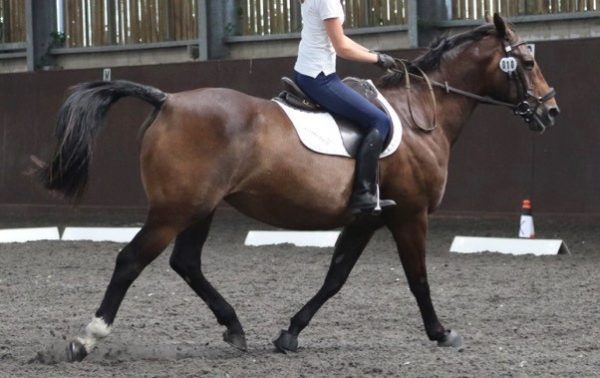
Head tilt, mouth open exposing teeth for 10 seconds, ears behind vertical for 5 seconds. Credit Dr Sue Dyson.
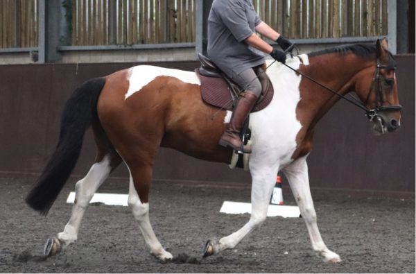
Ears behind vertical for 5 seconds; intense stare; mouth open exposing the teeth for 10 seconds; hindlimb toe drag. Credit Dr Sue Dyson.


